Upper Back Anatomy Organs : Thoracic Spine Major Muscles Physiopedia : This wall chart for understanding lower back pain has detailed illustrations of common back problems including herniated disks, scoliosis, osteoporosis.
Upper Back Anatomy Organs : Thoracic Spine Major Muscles Physiopedia : This wall chart for understanding lower back pain has detailed illustrations of common back problems including herniated disks, scoliosis, osteoporosis.. The sections below will cover these elements in more detail. Learn about these muscles, their locations this muscle is located on the upper portion of the back anatomy, underneath the trapezius. This bone contributes to the hard palate and holds the upper teeth. The final chapter presents anatomical charts of anatomical sections of the upper limb: The axilla and the deltoid region in axial and coronal and axial sections of the arm, the elbow, forearm, wrist, carpal and metacarpal regions.
Upper back pain and chest pain can occur together. Development of the human organism. The chest and upper back are in close proximity to each other with both sharing many ribs that help protect the same vital internal organs. This vein, as well as the deep veins. The anatomical areas found on the upper limb can serve as key landmarks to help us find important anatomical structures such as finding one of the superficial veins:
Solved Source Muscular System Views View 14 Upper Back Chegg Com from media.cheggcdn.com Instant anatomy is a specialised web site for you to learn all about human anatomy of the body with diagrams, podcasts and revision questions. Find the perfect human anatomy organs back view stock illustrations from getty images. Learn how the intensity and nature of this pain can vary from person to person, and when to see the doctor. The cervical spine is the top part of the spine. It could also refer to the sum total of organs and parts of one's body. Wolters kluwer health/lippincott anatomy and human movement: Musculoskeletal anatomy, kinesiology, and palpation for manual therapists. Structure and function (6th ed.).
This bone also contains a paranasal sinus that can be infected.
Structure and function (6th ed.). This bone also contains a paranasal sinus that can be infected. This wall chart for understanding lower back pain has detailed illustrations of common back problems including herniated disks, scoliosis, osteoporosis. Vertebrae help your body carry its weight efficiently, in addition to protecting the spinal cord and internal organs. During the 23rd century, most physicians had little or no familiarity with klingon anatomy. They're some of the most complex and frequently used this refers to moving a body part toward the center of the body, such as bringing the arm back in so it. Besides arm anatomy, we'll also teach you about some common conditions that can affect the arm the arms are the upper limbs of the body. It could also refer to the sum total of organs and parts of one's body. The back comprises the spine and spinal nerves, as well as several different muscle groups. Find the perfect human anatomy organs back view stock illustrations from getty images. The organs aren't really on your back they are situated between the framework of your body on the right side you have the the right lung, the liver, the gall bladder sections of your small and large intestine, your diaphragm, your right kidney, your appendix, the aorta runs midway through on the left. The nervous system of the thorax is a vital part of the nervous system as a whole, as it includes the spinal cord, peripheral nerves, and autonomic ganglia that communicate with and control many vital organs. Wolters kluwer health/lippincott anatomy and human movement:
Superficial (move shoulder and upper ext) and intermediate (mo… adducts, retracts, elevates/depresses scapula. This bone contributes to the hard palate and holds the upper teeth. Learn about back anatomy with free interactive flashcards. This bone also contains a paranasal sinus that can be infected. Vertebrae help your body carry its weight efficiently, in addition to protecting the spinal cord and internal organs.
Are You Suffering From Neck Shoulder Or Upper Back Pain The Center For Health And Sports Medicine from jaxfamilysportsmed.com These bones form the upper jaw. In the rear of the abdomen are the back muscles and spine. It could also refer to the sum total of organs and parts of one's body. During the 23rd century, most physicians had little or no familiarity with klingon anatomy. Besides arm anatomy, we'll also teach you about some common conditions that can affect the arm the arms are the upper limbs of the body. Where it joints the parietal bones anteriorly it forms the lambdoid suture. The final chapter presents anatomical charts of anatomical sections of the upper limb: Before one can understand how xenobiotics affect these different body an organ is a unique anatomic structure consisting of groups of tissues that work in concert to perform specific use the menu button in the upper right corner of each page to access the main sections.
Their main function is exchanging oxygen and.
The cervical spine is the top part of the spine. This bone contributes to the hard palate and holds the upper teeth. Standard anatomical position is the body orientation used when describing an organism's anatomy. Where it joints the parietal bones anteriorly it forms the lambdoid suture. Upper back pain and chest pain can occur together. A collection of articles covering upper limb anatomy topics, including the brachial plexus, bones of the hand and more. Learn about back anatomy with free interactive flashcards. Vertebrae help your body carry its weight efficiently, in addition to protecting the spinal cord and internal organs. The sections below will cover these elements in more detail. Development of the human organism. The cervical spine supports the weight and movement of your head and protects the nerves exiting your brain. Superficial (move shoulder and upper ext) and intermediate (mo… adducts, retracts, elevates/depresses scapula. Conditions › back pain › upper back pain › anatomy of the upper back.
Thoracic vertebrae have a few unique characteristics, as demonstrated in the illustration below. Upper back pain and chest pain can occur together. The sections below will cover these elements in more detail. Topographically, the muscles in this group are classed along with the lateral torso wall and upper extremity, which is due to their location as well as their genetic development based on their embryological origin. The cervical spine is the top part of the spine.
Trapezius Muscle Anatomy And Function from www.verywellhealth.com The back anatomy includes the latissimus dorsi, trapezius, erector spinae, rhomboid, & teres major. The organs aren't really on your back they are situated between the framework of your body on the right side you have the the right lung, the liver, the gall bladder sections of your small and large intestine, your diaphragm, your right kidney, your appendix, the aorta runs midway through on the left. Anatomy,back bone,back view,bone,bone structure,bones,bones of t, medical image collection, 87396753 10x8 (25x20cm) print neck muscle anatomy body anatomy anatomy study anatomy reference anatomy models anatomy for artists skeleton muscles thoracic vertebrae upper back. Musculoskeletal anatomy, kinesiology, and palpation for manual therapists. Wolters kluwer health/lippincott anatomy and human movement: The nervous system of the thorax is a vital part of the nervous system as a whole, as it includes the spinal cord, peripheral nerves, and autonomic ganglia that communicate with and control many vital organs. Superficial (move shoulder and upper ext) and intermediate (mo… adducts, retracts, elevates/depresses scapula. The median cubital vein (a common site site for venepuncture) in the antecubital fossa of the arm.
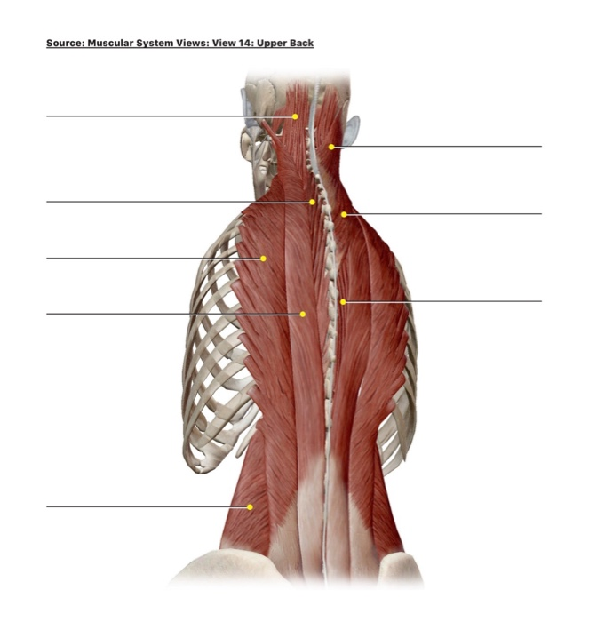
In the upper back region, the trapezius, rhomboid major, and levator scapulae muscles anchor the scapula and clavicle to the spines of several vertebrae and the occipital bone of the skull.
Structure and function (6th ed.). Before one can understand how xenobiotics affect these different body an organ is a unique anatomic structure consisting of groups of tissues that work in concert to perform specific use the menu button in the upper right corner of each page to access the main sections. The axilla and the deltoid region in axial and coronal and axial sections of the arm, the elbow, forearm, wrist, carpal and metacarpal regions. … the two kidneys are located in the back of the abdomen on either side of the body. The final chapter presents anatomical charts of anatomical sections of the upper limb: These organs are held together loosely by connecting tissues (mesentery) that allow them to expand and to slide against each other. The extrinsic back muscles are also referred to as secondary back muscles. Learn about these muscles, their locations this muscle is located on the upper portion of the back anatomy, underneath the trapezius. Organs exist in most multicellular organisms, including not only humans and other animals but also plants. Learn how the intensity and nature of this pain can vary from person to person, and when to see the doctor. Wolters kluwer health/lippincott anatomy and human movement: The back comprises the spine and spinal nerves, as well as several different muscle groups. The cervical spine supports the weight and movement of your head and protects the nerves exiting your brain.
The occipital bone forms the floor and the back wall of the skull upper back anatomy. This bone contributes to the hard palate and holds the upper teeth.

/GettyImages-147219941-56a05efe3df78cafdaa14c5f.jpg)